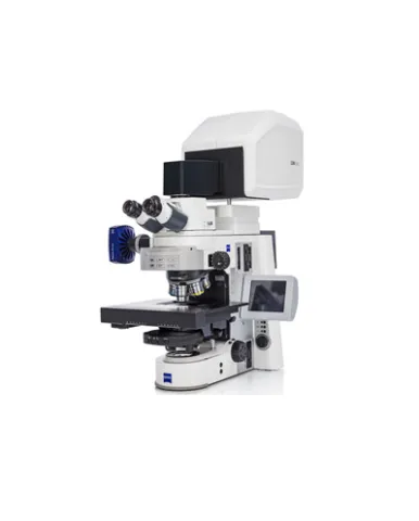
ZEISS LSM 900, the confocal laser scanning microscope (CLSM) from ZEISS, is the one instrument you will need for materials analysis. Characterize 3D microstructures surfaces topography in your lab or multi-user facility. Combine all essential light microscopy contrasting techniques for materials with high precision topography. Save time on set-up as there is no need to change microscopes. Execute non-contact confocal imaging when evaluating surface roughness. LSM 900 is the ideal tool for a multi-user facility. Extend your upright light microscope, ZEISS Axio Imager.Z2m or your inverted light microscope ZEISS Axio Observer 7, with a confocal scanning module.
Spesifikasi: Stands: Upright: Axio Imager.Z2m; Inverted: Axio Observer 7 z drive: Smallest increment 10 nm XY Stage (option): Motorized XY scanning stage for Mark & Find function (xy) as well as Tile Scan (Mosaic Scan). Tiling not available for layer thickness measurements. Smallest increment 0.2 μm Objectives: More than 40 reflected-light objectives. Recommended: C Epiplan-APOCHROMAT series (specially designed for 405 nm) Scanner: Two independent, galvanometric scanning mirrors with ultra-short line and frame flyback Scanning Resolution: 32 × 1 to 6,144 × 6,144 pixels, continuously adjustable (for each axis) Scanning Speed: Up to 8 images/sec with 1024 × 256 pixels; Up to 2 images/sec with 1024 × 1024 pixels Scanning Zoom: 0.5× to 40×; continuously adjustable Scanning Rotation: Can be rotated freely (360°), adjustable in increments of 0.1°, freely adjustable xy offset Scanning Field: 12.7 mm × 12.7 mm in the intermediate image plane with full pupil illumination Pinhole: Master pinhole with preset size and position; automatic alignment Beam Path: One major beam splitter at 10 degrees provides an 80:20 split ratio in a single-channel system. In multi-channel-systems there is an 80:20 split ratio at 405 nm and excellent laser line suppression at 488, 561 and 640 nm. The patented Variable Secondary Dichroics (VSDs) can be used as a flexible means of diverting the respective spectral range of light to the chosen channels. Use emission filters to clean up the signal when imaging autofluorescent or highly scattering samples. Detectors: Depending on whether it is configured with 1 or 2 multi-alkali (MA) PMT (typical QE25%) 2-CH scanhead can be further upgrated by 1 additional GaAsP PMT (typical QE 45%), MA PMT or Airyscan detector Transmitted light detector (ESID or T-PMT) Data Depth: 8-bit and 16-bit available Real-Time Electronics: Microscope, laser, scanning module and additional accessory control; data acquisition and synchronization management through real-time electronics; oversampling read-out logic for best sensitivity; data transfer between real-time electronics and user PC via LVDS with the ability to evaluate the data online during image acquisition
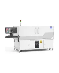
ZEISS Xradia CrystalCT® computed tomography platform uniquely augments this powerful imaging technique with the ability to reveal crystallographic grain microstructures, transforming the way polycrystalline materials (such as metals, additive manufacturing, ceramics, pharmaceuticals and others) can be studied, leading to newer and deeper insights for your materials research.
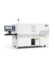
ZEISS Xradia Context® micro-computed tomography (microCT) is an easy-to-use system for analysis of all types of samples. A high-array detector enables high resolution of fine details even with relatively large imaging volumes. The system features a large field of view, rapid sample mounting and alignment, streamlined acquisition workflow and fast exposure and data reconstruction times.
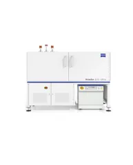
In XRM, contrast depends on the material being imaged and the X-ray energy used. The Xradia Ultra family comprises of Xradia 800 Ultra, operating at 8 keV photon energy, and Xradia 810 Ultra, operating at 5.4 keV. In general, lower energy X-rays are absorbed more strongly and therefore will provide you with higher contrast for most materials. Thus, as long as transmission remains sufficient, you will experience resulting image quality and/or throughput that are greatly improved with Xradia 810 Ultra.