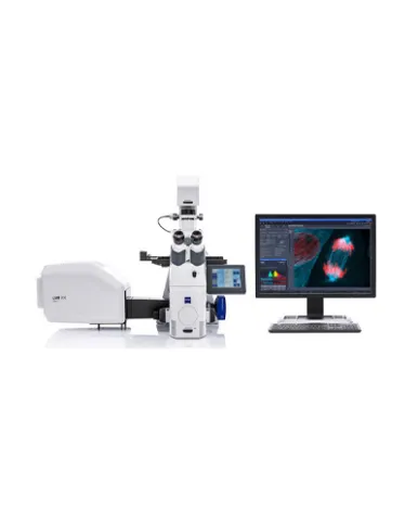
To pursue your scientific questions, you want only the best data quality. In microscopy, this translates into the best contrast and resolution while maintaining minimum light exposure. LSM 900, your compact confocal microscope, provides this with components optimized to deliver the best imaging results.
Spesifikasi: Scanner: Two independent, galvanometric scanning mirrors with ultrashort line and frame flyback Scanning resolution: 32 × 1 to 6,144 × 6,144 pixels (Airyscan 2 max. 4,096 × 4,096 pixels), also for multiple channels, continuously adjustable (for each axis) Scanning speed: At 512 × 512 pixels: confocal – up to 8 fps; Airyscan SR – up to 4 fps; Multiplex SR-2Y – 8.4 fps; Multiplex SR-4Y – 18.9 fps At 512 × 64 pixels: confocal – up to 64 fps Scanning zoom: 0.45 × to 40 ×; continuously adjustable Scanning rotation: Can be rotated freely (360°), adjustable in increments of 0.1°, freely adjustable xy offset Scanning field: 20 mm diagonal in the intermediate image plane, with full pupil illumination Pinhole: Master pinhole with preset size and position; can be adjusted as desired for multitracking and short wavelengths (such as 405 nm); automatic alignment Beam path: One major beam splitter for four laser lines (405, 488, 561 and 640 nm) at 10 degree with excellent laser line suppression. The 640 nm laser line can be used for internal autofocusing. Depending on the system, either one or two patented Variable Secondary Dichroics (VSD) can be used to flexibly divert the respective spectral range of light to chosen channels. Emission filters can be used to clean up the signal when imaging autofluorescent or highly scattering samples Physical Dimensions Length (cm) Width (cm) Height (cm) Weight (kg): Small actively and passively damped system table: 90 - 75 - 83 - 130 Large actively damped system table (incl. corner pieces): 120 (129) - 90 (99) - 87 - 180 Vibraplate for Axio Imager (consists of three pedestals): 32 - 30 - 4.5 - 1.5 Detectors: 2 spectral detection channels, GaAsP (typical QE 45 %) or multialkali (MA) PMT (typical QE 25 %); LSM Plus: resolution down to 160* nm lateral, 500 nm axial with pinhole at 0.8 AU; resolution down to 120* nm lateral, 500 nm axial with pinhole at 0.3 AU 1 additional GaAsP PMT, MA PMT or Airyscan 2 detector Airyscan 2 for spatial detection (GaAsP) with 40×, or 63×, or 100× objectives; for super-resolution (down to 120* nm lateral, 350 nm axial; with jDCV 90* nm lateral, 270 nm axial) or Multiplex acquisition (down to 140 nm) Transmitted light detector (ESID or T-PMT); unique transmitted fluorescence Sample Navigation with T-PMT Spectral detection: > 8 sequential confocal fluorescence channels, up to three parallel confocal fluorescence channels, based on low-noise GaAsP or MA PMTs; adjustable in increments of 1 nm Data depth: 8-bit and 16-bit available Real-time electronics: Microscope, laser, scanning module and additional accessory control; data acquisition and synchronization management through real-time electronics; oversampling read-out logic for best sensitivity; data transfer between real-time electronics and user PC via LVDS with the ability to evaluate the data online during image acquisition
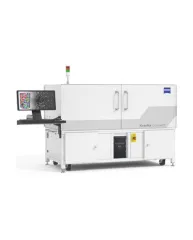
ZEISS Xradia CrystalCT® computed tomography platform uniquely augments this powerful imaging technique with the ability to reveal crystallographic grain microstructures, transforming the way polycrystalline materials (such as metals, additive manufacturing, ceramics, pharmaceuticals and others) can be studied, leading to newer and deeper insights for your materials research.
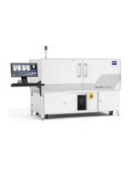
ZEISS Xradia Context® micro-computed tomography (microCT) is an easy-to-use system for analysis of all types of samples. A high-array detector enables high resolution of fine details even with relatively large imaging volumes. The system features a large field of view, rapid sample mounting and alignment, streamlined acquisition workflow and fast exposure and data reconstruction times.
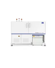
In XRM, contrast depends on the material being imaged and the X-ray energy used. The Xradia Ultra family comprises of Xradia 800 Ultra, operating at 8 keV photon energy, and Xradia 810 Ultra, operating at 5.4 keV. In general, lower energy X-rays are absorbed more strongly and therefore will provide you with higher contrast for most materials. Thus, as long as transmission remains sufficient, you will experience resulting image quality and/or throughput that are greatly improved with Xradia 810 Ultra.