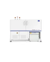
ZEISS Lattice Lightsheet 7 makes light sheet fluorescence microscopy available for live cell imaging at subcellular resolution – while also allowing you to use your standard sample carriers. With this automated, easy-to-use system, volumetric imaging of subcellular structures and dynamics over hours and days with best protection from photo damage becomes available to everyone. Discover the dynamics of life in unprecedented depth of detail – with the ease you never imagined possible!
Spesifikasi: Core Optics: Illumination objective lens 13.3× / 0.4 (at 30° angle to cover glass) with static phase element Detection objective lens 44.83× / 1.0 (at 60° angle to cover glass) with Alvarez manipulator Meniscus lens; relay of core optics to cover slip of sample carrier Autoimmersion: water, motorized dispenser Illumination: Transmitted light: LED (white centered and side, red centered illumination) with oblique contrast for sample positioning and overview; no Köhler Illumination, not specified for high quality imaging Reflected light and epifluorescence: laser (488 nm, 561 nm, 640 nm) for beam adjustment and fluorescence imaging Detection Modules: Hamamatsu ORCA-Fusion sCMOS camera; requires liquid cooling; up to two camera ports (right side port: camera 1, rear port: camera 2) Pixel size: 6.5 µm; Max. pixel format: 2,048 × 2,048 (4.2 Megapixel); Bit depth: 16 bit; QE: up to 80% Image Acquisition Speed: Volume: 3 Vol/s @ approx. 300 µm × 50 µm × 20 µm Plane: 400 frames/s @ approx. 300 µm × 20 µm Up to 3 colors fast sequential (framewise or stackwise switch) System PC / Workstation: HP Z6 G4 Rev2 workstation Chipset: Intel C622 Memory: max. 192 GB RAM SSD: 1× 512 GB M.2 NVMe (for pagefile and operating system); 1× MTE662T2 M.2 PCIe NVMe 2 TB Hard Drives: 2× 6 TB SATA 7,200 rpm (configured as 6 TB RAID 10 hard drive); increase capacity from 6 TB (RAID 10) to 12 TB (RAID 10) Processor: Intel® Xeon® Gold 6234 (3.2 GHz, 24.75 MB cache, 8 cores) Graphics Card: NVIDIA Quadro RTX6000 24 GB DB Network Adapter: 2× 10GbE RJ45 (hp Z6); additional network adapter 2× 10GbE RJ45 (hp Z6) e.g. for connection of storage systems Operating system: Windows 10 IoT Enterprise 2019 LTSC Embedded ×64 Main System Module Lightsheet: 7 600 mm × 425 mm × 380 mm 48 kg Component Rack (houses Laser module,Power supply unit & Stage Motion Controller): 550 mm × 740 mm × 600 mm 56 kg System Table for main System Module Lattice Lightsheet 7, Level regulated: 900 mm × 750 mm × 830 mm 130 kg Penetration Depth: Up to 200 µm Field of View (FOV): x: 300 µm; y: defined by scan range Spectral Range of Detection: 490 nm – 740 nm
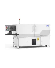
ZEISS Xradia CrystalCT® computed tomography platform uniquely augments this powerful imaging technique with the ability to reveal crystallographic grain microstructures, transforming the way polycrystalline materials (such as metals, additive manufacturing, ceramics, pharmaceuticals and others) can be studied, leading to newer and deeper insights for your materials research.
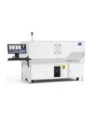
ZEISS Xradia Context® micro-computed tomography (microCT) is an easy-to-use system for analysis of all types of samples. A high-array detector enables high resolution of fine details even with relatively large imaging volumes. The system features a large field of view, rapid sample mounting and alignment, streamlined acquisition workflow and fast exposure and data reconstruction times.
In XRM, contrast depends on the material being imaged and the X-ray energy used. The Xradia Ultra family comprises of Xradia 800 Ultra, operating at 8 keV photon energy, and Xradia 810 Ultra, operating at 5.4 keV. In general, lower energy X-rays are absorbed more strongly and therefore will provide you with higher contrast for most materials. Thus, as long as transmission remains sufficient, you will experience resulting image quality and/or throughput that are greatly improved with Xradia 810 Ultra.