

Product
More From Microscope
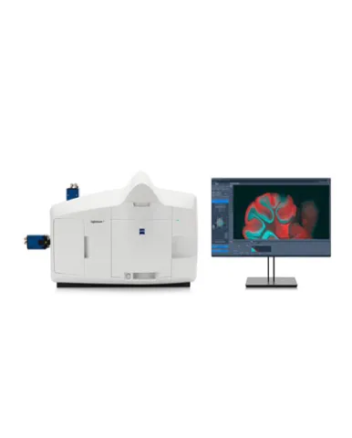
Xray Microscopy - ZEISS Xradia CrystalCT
ZEISS Xradia CrystalCT® computed tomography platform uniquely augments this powerful imaging technique with the ability to reveal crystallographic grain microstructures, transforming the way polycrystalline materials (such as metals, additive manufacturing, ceramics, pharmaceuticals and others) can be studied, leading to newer and deeper insights for your materials research.
Rp.123
See Details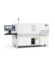
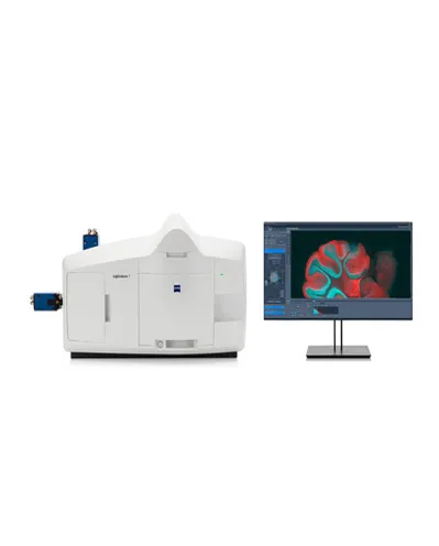
Xray Microscopy - ZEISS Xradia Context MicroCT
ZEISS Xradia Context® micro-computed tomography (microCT) is an easy-to-use system for analysis of all types of samples. A high-array detector enables high resolution of fine details even with relatively large imaging volumes. The system features a large field of view, rapid sample mounting and alignment, streamlined acquisition workflow and fast exposure and data reconstruction times.
Rp.123
See Details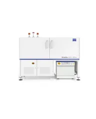
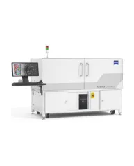
Xradia Ultra - ZEISS Xradia 810 or 800 Ultra
In XRM, contrast depends on the material being imaged and the X-ray energy used. The Xradia Ultra family comprises of Xradia 800 Ultra, operating at 8 keV photon energy, and Xradia 810 Ultra, operating at 5.4 keV. In general, lower energy X-rays are absorbed more strongly and therefore will provide you with higher contrast for most materials. Thus, as long as transmission remains sufficient, you will experience resulting image quality and/or throughput that are greatly improved with Xradia 810 Ultra.
Rp.123
See Details