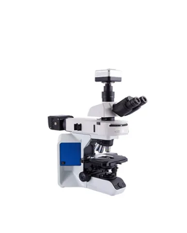
MJ43 is a research level metallurgical microscope with reflected lighting and transmitted lighting. Multi-function lighting attachment and broad-spectrum LED light source allows the microscope expand to reflected fluorescence function. The microscope body and structure is equivalent to Olympus BX43 optimized optical performance, offering various observation method possibility of dark field and DIC. All features give the microscope system ability to achieve composite micro observation of semi-conductor, FPD, electronic board, precision metal parts, physical materials and related industry area. It’s an ideal instrument for university laboratory, education and scientific research purpose.
Spesifikasi: Observation: Hinged type trinocular observation tube with 30°inclination Wide field eyepiece 10X/22mm, high eye-point, interpupillary distance 53~75mm Nosepiece: Quintuple nosepiece Objectives Semi-apochromatic metallurgical objective PlanFluor EPI 5X/0.15,WD:20mm Semi-apochromatic metallurgical objective PlanFluor EPI 10X/0.3,WD:11mm Semi-apochromatic metallurgical objective PlanFluor EPI 20X/0.45,WD:3mm Semi-apochromatic metallurgical objective PlanFluor EPI 50X/0.8,WD:1mm Stage: Coaxial stage with left or right hand low drive control: with rotating mechanism and torque adjustment mechanism Travel distance 80mm×50mm, 1mm per scale, accuracy 0.1mm Triple-layer mechanical stage with glass plate clamp Transparent glass plate 97mm×76mm Reflected illumination: Turret accommodates maximum sextuple filter block cubes Mirror units of bright field Digital control LED light control box, AC110V/220V 30W board-spectrum LED light source MG30 Connector of broad-spectrum LED light source Analyzer and polarizer Transmitted illumination: system Warm white LED lamp, brightness is continuously adjustable Abbe condenser, N.A. 1.1 Power line: According to user's country electrical requirement Screw driver: M3.0 hexagon screw driver Accessories: Immersion oil Dust cover: MSHOT special dust cover Optional adapters: 1X/0.5X/0.75X C-mount adapter
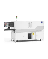
ZEISS Xradia CrystalCT® computed tomography platform uniquely augments this powerful imaging technique with the ability to reveal crystallographic grain microstructures, transforming the way polycrystalline materials (such as metals, additive manufacturing, ceramics, pharmaceuticals and others) can be studied, leading to newer and deeper insights for your materials research.
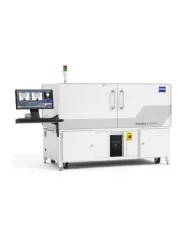
ZEISS Xradia Context® micro-computed tomography (microCT) is an easy-to-use system for analysis of all types of samples. A high-array detector enables high resolution of fine details even with relatively large imaging volumes. The system features a large field of view, rapid sample mounting and alignment, streamlined acquisition workflow and fast exposure and data reconstruction times.
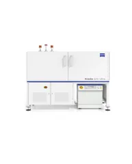
In XRM, contrast depends on the material being imaged and the X-ray energy used. The Xradia Ultra family comprises of Xradia 800 Ultra, operating at 8 keV photon energy, and Xradia 810 Ultra, operating at 5.4 keV. In general, lower energy X-rays are absorbed more strongly and therefore will provide you with higher contrast for most materials. Thus, as long as transmission remains sufficient, you will experience resulting image quality and/or throughput that are greatly improved with Xradia 810 Ultra.