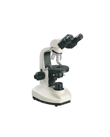
MP30 Polarizing Microscope is a kind of essential instrument for studying and identifying the materials with double refraction characteristics, by using polarizing feature. This microscope is widely used in many fields, such as observation of crystalline inclusions, muscle tissue, nerve tissue, plant fiber and other structural details of the teeth, bone, and living cells, as well as analysis of drug composition and properties. Furthermore, in the field of inorganic chemistry, the fine structure of crystallization conditions of various kinds of salts which cannot be seen in natural light can be observation through polarizing microscope.
Spesifikasi: Model: MP30 Observation tube: Trinocular, inclined 30°, 100% light pass for photography capable Eyepiece: Wide field WF10X(Φ22mm) Graduated WF10X(Φ22mm) 0.10mm/Div Objective: Strain-free plan achromatic objective (no cover glass) PL L5X/0.12 (Work distance):26.1 mm PL L10X/0.25 (Work distance):20.2 mm PL L40X/0.60(spring) (Work distance):3.98 mm PL L60X/0.70(spring) (Work distance):3.18 mm Reflective illuminating system: 6V30W halogen, brightness adjustable. Polarizer can be rotated 360°. Analyzer can be rotatable 360° with scale and minimum vernier Integrated field diaphragm and aperture diaphragm Nosepiece:Quadruple (center adjustable ) Intermediate attachments: Pull type Bertrand lens and center adjustable Compensator λ ,λ/4 and quarts wedge compensator Focus system: Coaxial coarse/fine focus system, with tension adjustable and limit stopper, Minimum graduation in fine movement: 2μm Transmitted illuminating system: 12V30W halogen, brightness adjustable, lamp center adjustable Abbey condenser,liftable,NA 1.25 Blue filter Frosted glass Polarizer (360° rotatable with 0°,90°,180°,270° division) Camera adapter: 1X C-mount adapter
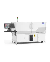
ZEISS Xradia CrystalCT® computed tomography platform uniquely augments this powerful imaging technique with the ability to reveal crystallographic grain microstructures, transforming the way polycrystalline materials (such as metals, additive manufacturing, ceramics, pharmaceuticals and others) can be studied, leading to newer and deeper insights for your materials research.
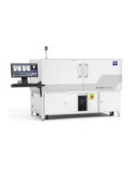
ZEISS Xradia Context® micro-computed tomography (microCT) is an easy-to-use system for analysis of all types of samples. A high-array detector enables high resolution of fine details even with relatively large imaging volumes. The system features a large field of view, rapid sample mounting and alignment, streamlined acquisition workflow and fast exposure and data reconstruction times.
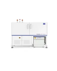
In XRM, contrast depends on the material being imaged and the X-ray energy used. The Xradia Ultra family comprises of Xradia 800 Ultra, operating at 8 keV photon energy, and Xradia 810 Ultra, operating at 5.4 keV. In general, lower energy X-rays are absorbed more strongly and therefore will provide you with higher contrast for most materials. Thus, as long as transmission remains sufficient, you will experience resulting image quality and/or throughput that are greatly improved with Xradia 810 Ultra.