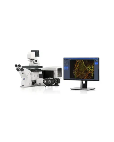
ZEISS Elyra 7 includes a wealth of microscopy techniques to meet your experimental needs across scales, optimally matching resolution, speed, and sensitivity requirements to your demanding samples. Employ SIM Apotome for fast optical sectioning, Lattice SIM for super-resolution imaging, SIM² image reconstruction for resolution excellence down to 60 nm, as well as SMLM and TIRF for investigations at the molecular level. You can combine these techniques to multiply the insights from your specimen and to correlate the acquired data.
Spesifikasi: Stand: Axio Observer 7, motorized inverted microscope for superresolution microscopy Z-drive: DC servomotor, opto-electronically coded; smallest Z step 25 nm XY Piezo scanning stage: motorized; range 130 mm× 100 mm; max sped 100 mm/s; resolution 0.2 µm; reproducibility: ± 1 µm; absolute accuracy ± 5 µm; suitable for mounting frames K 160 × 110 mm and Z-Piezo Stage insert Z-Piezo stage insert: for XY scanning stage, max travel range 500 µm; smallest Z step size 5 nm, sample holders available for standard 3"×1" slides LabTek chambers, multiwell plates and 36 mm glass-bottom dishes; level-adjustable and universal stage insert available for standard slides, glass-bottom dishes and LabTekTM chambers. Filter sets reflector turret: Four exchangeable filter sets available for multi-channel Lattice SIM and SMLM; each filter set with four precisely mounted ACR-coded(1) filter modules for superresolution microscopy on a motorized six-position turret; two positions in each turret compatible with standard Push & Click filter modules, e.g. for visual sample observation. Dual filter sets for Duolink optimized for dual color and double dual color applications: Filter sets are optimized for dual camera applications, maximum sensitivity, minimal cross-talk and reduced autofluorescence. Filter slider: Manual filter slider with two positions (for emission filters or a Bertrand lens); fits into camera adapter of the microscope’s side port; emission filters exchangeable for customizing detection conditions. Laser module for Elyra 7: Laser coupling with polarization-maintaining single mode fiber (no adjustment of laser coupling by users required) Laser lines: 405 nm (50 mW), 488 nm (100 mW or 500 mW), 561 nm (100 mW or 500 mW), 642 nm (100 mW) or 640 nm (500 mW); 405 laser can be attenuated by up to 100000 fold (used for activation and back-pumping); high power lasers (500 mW) can be 10 fold attenuated (488, 561, 642) Localization precision (SMLM): Typically 10 nm – 20 nm lateral, 20 nm – 40 nm axial, given sufficient signal-to-noise & density Multi-color imaging (SMLM): Detection of up to two different fluorescent labels (simultaneous with Duolink or quasi simultaneously by fast sequential laser switching) Acquisition speed (SMLM): sCMOS (dSTORM) and widefield mode > 100 frames per second full chip; 200 frames per second (512 × 512 pixels) Multi-color (Lattice SIM and SIM Apotome mode): Detection of up to four different fluorescent labels (sequential detection) and simultaneous dual color detection with DuoLink Max. Field of view (Lattice SIM): 80.14 × 80.14 μm², full-frame recording (1280 × 1280 effective px) with Plan-Apochromat 63× / 1.40 Oil DIC*
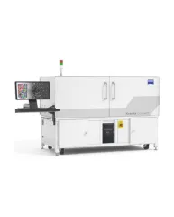
ZEISS Xradia CrystalCT® computed tomography platform uniquely augments this powerful imaging technique with the ability to reveal crystallographic grain microstructures, transforming the way polycrystalline materials (such as metals, additive manufacturing, ceramics, pharmaceuticals and others) can be studied, leading to newer and deeper insights for your materials research.
ZEISS Xradia Context® micro-computed tomography (microCT) is an easy-to-use system for analysis of all types of samples. A high-array detector enables high resolution of fine details even with relatively large imaging volumes. The system features a large field of view, rapid sample mounting and alignment, streamlined acquisition workflow and fast exposure and data reconstruction times.
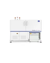
In XRM, contrast depends on the material being imaged and the X-ray energy used. The Xradia Ultra family comprises of Xradia 800 Ultra, operating at 8 keV photon energy, and Xradia 810 Ultra, operating at 5.4 keV. In general, lower energy X-rays are absorbed more strongly and therefore will provide you with higher contrast for most materials. Thus, as long as transmission remains sufficient, you will experience resulting image quality and/or throughput that are greatly improved with Xradia 810 Ultra.