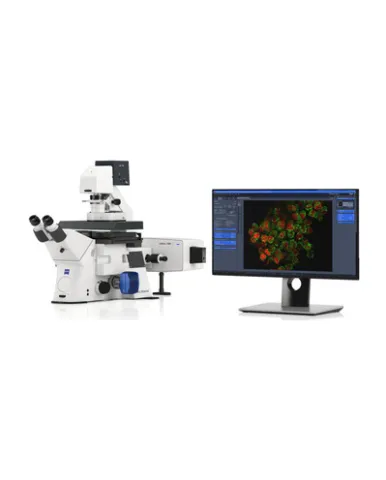
ZEISS Lattice SIM 3 is specifically designed to meet the imaging requirements of multicellular organisms and tissue sections. This system exploits the full potential of the SIM Apotome technology: fast optical sectioning at superior quality, large fields of view with access to smaller regions of interest, near-isotropic resolution, and the gentlest super-resolution imaging possible.
Spesifikasi: Stand: ZEISS Axio Observer 7, SR for Lattice SIM, motorized inverted microscope for super-resolution microscopy Z-drive: DC servo motor, opto-electronically coded; smallest Z step: 25 nm XY stepper scanning stage: Motorized, stepper motor with 2 mm spindle pitch; travel range: 130 mm × 100 mm; max. speed: 50 mm/s; Resolution: 0.1 µm; reproducibility: ± 1 µm; absolute accuracy: ± 5 µm; Suitable for mounting frames K 160 × 110 mm and Z-Piezo stage insert; compatible with objectives‘ autocorr Z-Piezo stage insert: For XY scanning stage; max travel range: 500 µm; smallest Z step size: 5 nm; Level-adjustable stage insert for frame inserts (sample holders) and multi-well plates; Sample holders available for 3“ × 1“ standard slides, LabTek chambers; 35 – 40 mm glass-bottom dishes; Universal stage insert: for various carrier formats Filter sets reflector turret Flexible filter set available for simultaneous multi-channel acquisition; Filter set with four precisely mounted ACR-coded filter modules for super-resolution microscopy on a motorized six-position turret; Two positions in the turret compatible with standard Push & Click filter modules, e.g., for visual sample observation Dual filter set for Duolink: Filter sets are optimized for one color (SOLO), dual color (DUO) and four color (QUAD) applications Filter slider: Manual filter slider with Bertrand lens; fits into the slit below the objective turret Laser module: Laser coupling with polarization-maintaining single mode fiber (no adjustment of laser coupling by users required) Laser lines: 405 nm (50 mW), 488 nm (50 mW), 561 nm (50 MW), 640 nm (50 mW); 405, 488 & 642 nm: diode lasers (DL); 561 nm: frequency doubled diode laser (FDDL); Direct modulation @ 500:1 Definite Focus: Holding focus to compensate axial drift, typical z-position accuracy: 30 nm; Specified limits of Definite Focus 3: 0.2 × DOF (Depth of field: DOF ≈ λ /NA²). Incubation: Stage top incubation with safety lock Duolink for attachment of two cameras of the same type: Allows attachment of two cameras of the same type to the microscope. Storage PC with 81 TByte storage capacity: Direct streaming of data and parallel processing while streaming of data possible
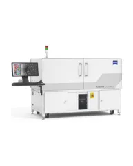
ZEISS Xradia CrystalCT® computed tomography platform uniquely augments this powerful imaging technique with the ability to reveal crystallographic grain microstructures, transforming the way polycrystalline materials (such as metals, additive manufacturing, ceramics, pharmaceuticals and others) can be studied, leading to newer and deeper insights for your materials research.
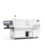
ZEISS Xradia Context® micro-computed tomography (microCT) is an easy-to-use system for analysis of all types of samples. A high-array detector enables high resolution of fine details even with relatively large imaging volumes. The system features a large field of view, rapid sample mounting and alignment, streamlined acquisition workflow and fast exposure and data reconstruction times.
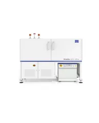
In XRM, contrast depends on the material being imaged and the X-ray energy used. The Xradia Ultra family comprises of Xradia 800 Ultra, operating at 8 keV photon energy, and Xradia 810 Ultra, operating at 5.4 keV. In general, lower energy X-rays are absorbed more strongly and therefore will provide you with higher contrast for most materials. Thus, as long as transmission remains sufficient, you will experience resulting image quality and/or throughput that are greatly improved with Xradia 810 Ultra.