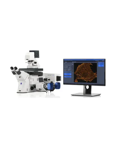
ZEISS Lattice SIM 5 has been optimized for capturing subcellular structures and their dynamics. Powered by the Lattice SIM technology and the SIM² image reconstruction algorithm, ZEISS Lattice SIM 5 provides you with outstanding super-resolution capabilities down to 60 nm in both living and fixed cells. Additionally, you can choose SIM Apotome imaging mode and a low-magnification objective to achieve fast overview images of your sample before zooming into super-resolution details.
Spesifikasi: Definite: Focus Holding focus to compensate axial drift, typical z-position accuracy: 30 nm; Specified limits of Definite Focus 3: 0.2 × DOF (Depth of field: DOF ≈ λ /NA²). Incubation: Stage top incubation with safety lock Duolink for attachment of two cameras of the same type: Allows attachment of two cameras of the same type to the microscope. Storage PC with 81 TByte storage capacity: Direct streaming of data and parallel processing while streaming of data possible Filter sets reflector turret: Flexible filter set available for simultaneous multi-channel acquisition Filter set with four precisely mounted ACR-coded filter modules for super-resolution microscopy on a motorized six-position turret; Two positions in the turret compatible with standard Push & Click filter modules, e.g., for visual sample observation Dual filter set for Duolink: Filter sets are optimized for one color (SOLO), dual color (DUO) and four color (QUAD) applications Filter slider: Manual filter slider with Bertrand lens; fits into the slit below the objective turret Laser module for Lattice SIM 5: Laser coupling with polarization-maintaining single mode fiber (no adjustment of laser coupling by users required) Laser lines: 405 nm (50 mW), 488 nm (50 mW), 561 nm (50 MW), 640 nm (50 mW); 405, 488 & 640 nm: diode lasers (DL); 561 nm: frequency doubled diode laser (FDDL); Direct modulation @ 500:1 Resolution (SIM/ SIM² Apotome): Lateral resolution (XY): down to 320 / 265 nm for 25× (typical experimental FWHM values with sub-resolution beads of 100 nm diameter and excitation at 488 nm) Multi-color (Lattice SIM and SIM Apotome): Detection of up to four different fluorescent labels (sequential detection) and simultaneous dual-color detection with Duolink Max. field of view (Lattice SIM) @ ORCA-Fusion BT: 103.21 × 103.21 μm², full-frame recording (1288 × 1288 effective px) with Plan-Apochromat 63× / 1.40 Oil DIC* Max. field of view (SIM Apotome) @ ORCA-Fusion BT: 127 × 127 µm², full frame recording (1288 × 1288 effective px) with Plan-Apochromat 40× / 1.40 Oil; 203.20 × 203.20 µm², full frame recording with LD LCI Plan-Apochromat 25× / 0.8 Imm Corr DIC*; 254 × 254 µm², full frame recording with Plan-Apochromat 20× / 0.8 Air; 651 × 651 µm², full frame recording with EC Plan-Neofluar 10× / 0.3 Air
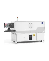
ZEISS Xradia CrystalCT® computed tomography platform uniquely augments this powerful imaging technique with the ability to reveal crystallographic grain microstructures, transforming the way polycrystalline materials (such as metals, additive manufacturing, ceramics, pharmaceuticals and others) can be studied, leading to newer and deeper insights for your materials research.
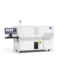
ZEISS Xradia Context® micro-computed tomography (microCT) is an easy-to-use system for analysis of all types of samples. A high-array detector enables high resolution of fine details even with relatively large imaging volumes. The system features a large field of view, rapid sample mounting and alignment, streamlined acquisition workflow and fast exposure and data reconstruction times.
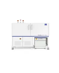
In XRM, contrast depends on the material being imaged and the X-ray energy used. The Xradia Ultra family comprises of Xradia 800 Ultra, operating at 8 keV photon energy, and Xradia 810 Ultra, operating at 5.4 keV. In general, lower energy X-rays are absorbed more strongly and therefore will provide you with higher contrast for most materials. Thus, as long as transmission remains sufficient, you will experience resulting image quality and/or throughput that are greatly improved with Xradia 810 Ultra.