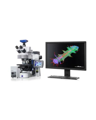
Combine the best for life science research in a single upright platform. Whether you are simply observing and recording or performing highly complex imaging experiments: It's easy to customize system components to meet the needs of your applications.
Spesifikasi: Axio Imager.A2 LED Standard Equipment: LED – Fixed-Koehler illumination transmitted-light Light manager Encoded Optional Equipment: Transmitted-light beam path with manual filter wheel Reflected-light beam path Apotome 3 Encoded stage Supported Applications: Evaluation Fast routine work Axio Imager.A2 Standard Equipment: Universal stand transmitted-light Light manager Encoded Neutral density filter wheel Optional Equipment: Reflected-light beam path Apotome 3 Encoded and 2-plate scanning stages Supported Applications: Observation Image acquisition and reporting Interactive measurements Axio Imager.D2 Standard Equipment: Universal stand transmitted-light Encoded Partly motorizable: reflector turret Optional Equipment: Reflected-light beam path Reflector turret 6x or 10x Apotome 3 Encoded and 2-plate scanning stages Supported Applications: Evaluation Image acquisition and reporting Semi-automatic measurements Axio Imager.M2p Standard Equipment: LED – Fixed-Koehler illumination transmitted-light Convenience motorization: par-focality, condenser Encoded nosepiece Motorized z-drive with 25 nm step size Optional Equipment Transmitted-light beam path with motorized luminous field stop Reflected-light beam path TFT Apotome 3 LSM (entry level) 2- and 3-plate scanning stages Supported Applications: Evaluation Image acquisition and reporting Fast routine work Axio Imager.M2 Standard Equipment: Universal stand transmitted-light Motorized: luminous field stop Light manager Contrast manager Motorized z-drive with 25nm step size Optional Equipment: Reflected-light beam path ACR for objectives Apotome 3 2- und 3-plate scanning stages 2 TV tube motorized Supported Applications: Automatic image acquisition & analysis 3D Imaging Medium sample throughput Multi-user environment Axio Imager.Z2 Standard Equipment: High-performance stand trans-mitted-light Motorized: luminous field stop Light manager Contrast manager Motorized focus drive: - 10 nm step size - designed for loads up to max. 9 kg - designed for continuous operation Optional Equipment: Reflected-light beam path ACR for objectives and filter cubes Apotome 3 2- and 3-plate scanning stages LSM Supported Applications: Automatic image acquisition & analysis Certified image acquisition and archiving (CFR 21 part 11) 3D imaging DIC fluorescence imaging Confocal imaging High sample throughput Multi-user environment
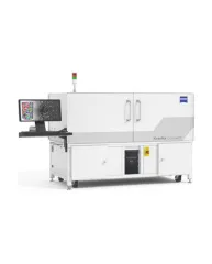
ZEISS Xradia CrystalCT® computed tomography platform uniquely augments this powerful imaging technique with the ability to reveal crystallographic grain microstructures, transforming the way polycrystalline materials (such as metals, additive manufacturing, ceramics, pharmaceuticals and others) can be studied, leading to newer and deeper insights for your materials research.
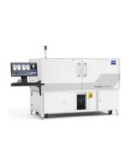
ZEISS Xradia Context® micro-computed tomography (microCT) is an easy-to-use system for analysis of all types of samples. A high-array detector enables high resolution of fine details even with relatively large imaging volumes. The system features a large field of view, rapid sample mounting and alignment, streamlined acquisition workflow and fast exposure and data reconstruction times.
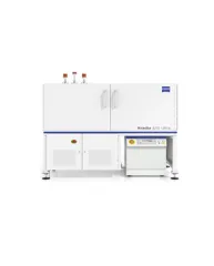
In XRM, contrast depends on the material being imaged and the X-ray energy used. The Xradia Ultra family comprises of Xradia 800 Ultra, operating at 8 keV photon energy, and Xradia 810 Ultra, operating at 5.4 keV. In general, lower energy X-rays are absorbed more strongly and therefore will provide you with higher contrast for most materials. Thus, as long as transmission remains sufficient, you will experience resulting image quality and/or throughput that are greatly improved with Xradia 810 Ultra.